Millions of Americans with diabetes are unknowingly putting their eyesight at risk — and a simple annual eye exam could change everything.
Introduction
Margaret, 54, had managed her type 2 diabetes for over a decade. She took her metformin faithfully, watched her carb intake, and showed up for most of her primary care appointments. But she hadn’t seen an eye doctor in four years — because her vision seemed just fine.
Then, almost overnight, she noticed blurry patches in her right eye. By the time she finally saw an ophthalmologist, she had moderate diabetic retinopathy in both eyes and was at serious risk of permanent vision loss.
Margaret’s story is not unusual. According to the CDC, diabetic retinopathy is the leading cause of new blindness among American adults aged 20 to 74. Yet research suggests that up to 90% of diabetes-related vision loss is preventable with timely screening and proper blood sugar management.
In this guide, you’ll learn exactly what diabetic retinopathy is, how it develops, what symptoms to watch for, and — most importantly — how you can protect your vision starting today.
What Is Diabetic Retinopathy?
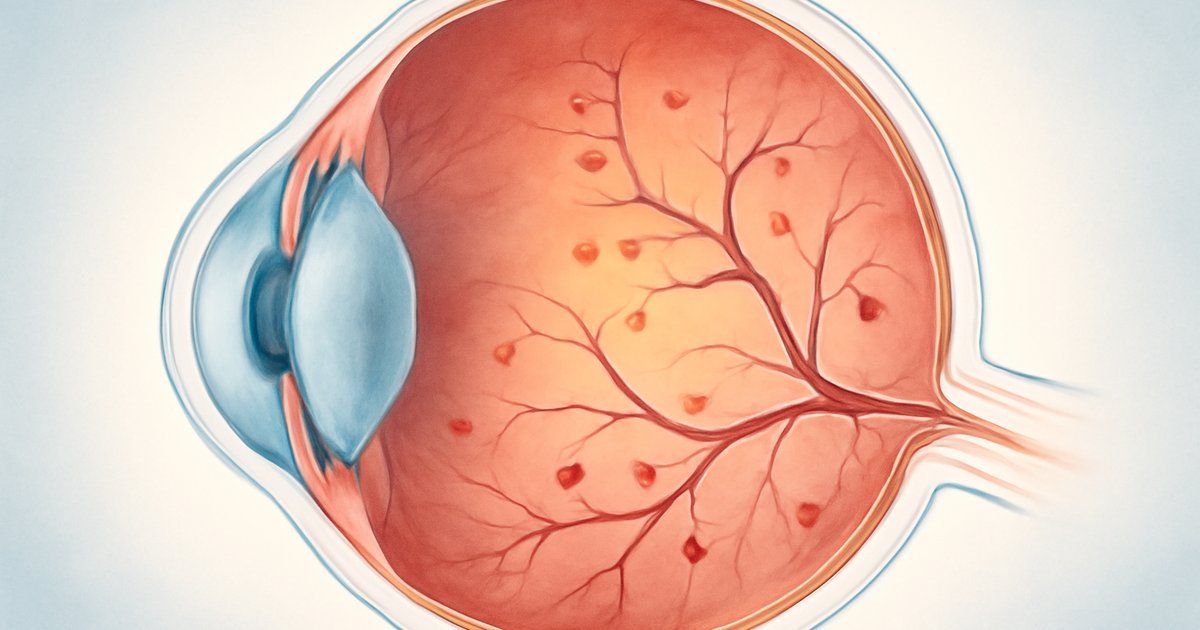
Diabetic retinopathy is a diabetes complication that damages the blood vessels in the retina — the thin, light-sensitive layer of tissue at the back of your eye. When blood sugar levels remain chronically elevated, those tiny vessels can swell, leak fluid, or grow abnormally, all of which can distort or destroy your vision over time.
The condition progresses in two main stages. Non-proliferative diabetic retinopathy (NPDR) is the earlier stage, where blood vessels in the retina weaken and may leak. Proliferative diabetic retinopathy (PDR) is the advanced stage, where new, abnormal blood vessels grow on the surface of the retina — a process called neovascularization — which dramatically increases the risk of severe vision loss or detachment of the retina.
According to the National Eye Institute (NEI), an arm of the NIH, approximately 7.7 million Americans have diabetic retinopathy, and that number is projected to reach nearly 14.6 million by 2050. Both type 1 and type 2 diabetes can cause the condition, though the longer you’ve lived with diabetes, the higher your risk becomes.
What makes diabetic retinopathy particularly dangerous is that it causes no pain and minimal early symptoms — meaning most people don’t realize anything is wrong until significant damage has already occurred.
Signs and Symptoms: What to Watch For
Because diabetic retinopathy often progresses silently, many people only notice symptoms once the disease has advanced. Understanding the full spectrum of warning signs — from subtle early changes to urgent red flags — can help you seek care at the right time.
Early signs (may be subtle or absent):
- Slightly blurry vision that comes and goes
- Difficulty reading fine print or seeing close objects
- Colors appearing washed out or less vibrant
- Small dark spots or "floaters" in your field of vision
- Difficulty seeing well in low-light conditions
Advanced signs (require urgent medical attention):
- Sudden or rapid vision loss in one or both eyes
- A large number of new floaters appearing suddenly
- Flashes of light in your vision
- A dark curtain or shadow appearing across part of your visual field
- Severe blurring that does not improve
A 2021 clinical study published through the American Academy of Ophthalmology found that nearly 40% of people with type 2 diabetes who were newly diagnosed with retinopathy reported having experienced no noticeable visual symptoms before their diagnosis. This underscores why routine eye exams are so critical — your eyes may be changing even when your vision feels normal.
Causes and Risk Factors
The root cause of diabetic retinopathy is sustained high blood sugar. Over time, excess glucose in the bloodstream damages the walls of the small blood vessels that supply the retina. These weakened vessels may swell, develop tiny bulges called microaneurysms, or leak blood and fluid into surrounding retinal tissue.
But not every person with diabetes develops retinopathy at the same rate or severity. Several factors can significantly increase your risk:
- Duration of diabetes: The longer you’ve had diabetes, the higher your cumulative risk. The NIH reports that nearly all people with type 1 diabetes and more than 60% of those with type 2 diabetes will develop some degree of retinopathy after 20 years.
- Poor blood sugar control: Chronically elevated A1C levels (a measure of average blood sugar over roughly three months) are one of the strongest predictors of retinopathy progression.
- High blood pressure (hypertension): Elevated blood pressure compounds damage to retinal blood vessels and is extremely common among people with diabetes.
- High cholesterol: Elevated LDL cholesterol increases the likelihood of hard exudates — fatty deposits — forming in the retina.
- Pregnancy: Gestational diabetes or pre-existing diabetes during pregnancy can accelerate retinopathy development.
- Smoking: Tobacco use constricts blood vessels and worsens microvascular complications throughout the body, including the eyes.
- Ethnicity: According to the CDC, African Americans, Hispanic/Latino Americans, and Native Americans face disproportionately higher rates of diabetic eye disease.
- Kidney disease: Diabetic nephropathy (kidney damage from diabetes) often occurs alongside retinopathy, as both stem from the same microvascular damage mechanisms.
It’s worth noting that individual variation plays a significant role here. Two people with identical A1C levels and the same duration of diabetes may experience very different rates of retinal damage — which is why personalized care with your healthcare team matters so much.
Diagnosis: What to Expect at the Eye Doctor
Diagnosing diabetic retinopathy requires a comprehensive dilated eye exam — a procedure in which your eye doctor uses special drops to widen (dilate) your pupils, giving them a clear view of your retina and the blood vessels within it. This exam is painless, though your vision may remain blurry for a few hours afterward.
Here’s what the diagnostic process typically involves:
- Visual acuity test: A standard eye chart test to measure how well you see at various distances.
- Dilated fundus examination: The doctor uses a special lens to examine the retina, optic nerve, and blood vessels directly.
- Optical coherence tomography (OCT): A non-invasive imaging scan that produces detailed cross-section images of the retina, allowing detection of swelling (macular edema) and structural changes.
- Fluorescein angiography: A dye is injected into the arm and photographs track how it flows through the retinal blood vessels, revealing leaks or abnormal growth areas.
The American Diabetes Association (ADA) recommends that adults with type 2 diabetes get a dilated eye exam at the time of diagnosis and annually thereafter. People with type 1 diabetes should begin annual exams within five years of diagnosis. If retinopathy is detected, your ophthalmologist may recommend more frequent monitoring — sometimes every three to six months.
If you’ve been skipping these exams, you’re far from alone. Research suggests that fewer than half of Americans with diabetes receive the recommended annual dilated eye exams. Consistent blood sugar monitoring goes hand in hand with eye exams as a cornerstone of diabetes management.
Treatment Options for Diabetic Retinopathy
The good news is that diabetic retinopathy responds well to treatment, especially when caught early. Your treatment plan will depend on the stage of the disease and whether diabetic macular edema (swelling at the center of the retina) is present.
1. Blood sugar and blood pressure management
For early-stage retinopathy, the most effective "treatment" is tighter control of blood glucose and blood pressure. Clinical evidence from the landmark DCCT (Diabetes Control and Complications Trial) and UKPDS (UK Prospective Diabetes Study) showed that intensive blood sugar control reduced the risk of retinopathy progression by 50–76% compared to conventional management. Most physicians recommend an A1C target below 7% for most adults with diabetes, though your personal target may vary.
2. Anti-VEGF injections
For moderate to severe retinopathy or diabetic macular edema, anti-VEGF (vascular endothelial growth factor) therapy is now the standard of care. Medications such as ranibizumab, aflibercept, and bevacizumab are injected directly into the eye to block the growth of abnormal blood vessels and reduce leakage. Clinical trials have shown these injections can significantly stabilize and even improve vision in many patients.
3. Laser photocoagulation therapy
Laser treatment has been used for decades to seal leaking blood vessels and shrink abnormal ones. Focal laser treatment targets specific leaking vessels, while panretinal photocoagulation (PRP) treats the peripheral retina to reduce the drive for abnormal vessel growth. While laser therapy is less commonly used as a first-line treatment since anti-VEGF injections became available, it remains an important option in many cases.
4. Vitrectomy surgery
In advanced cases where there is bleeding into the vitreous (the gel-like substance filling the eye) or a retinal detachment, a surgical procedure called vitrectomy may be necessary. The surgeon removes the vitreous and any scar tissue, and in some cases reattaches the retina. According to the NIH, vitrectomy significantly improves visual outcomes in patients with severe proliferative diabetic retinopathy when performed in a timely manner.
5. Lifestyle modifications as complements
While not a replacement for medical treatment, lifestyle changes provide meaningful support for retinopathy management. Regular physical activity, a diet low in refined carbohydrates and high in antioxidant-rich vegetables, smoking cessation, and stress reduction all contribute to better blood sugar control and vascular health. Emerging research also suggests that omega-3 fatty acids and certain antioxidant nutrients like lutein and zeaxanthin may support retinal health, though clinical evidence is still evolving.
Living With Diabetic Retinopathy and Preventing Progression
A diagnosis of diabetic retinopathy doesn’t have to mean inevitable vision loss. With proactive management and the right support team, many people successfully preserve their vision for years — and in some cases, decades.
Here are practical, evidence-informed strategies for day-to-day management:
- Keep your A1C in your target range. Work with your doctor to find the right A1C goal for your individual situation. Even a modest reduction in A1C — say, from 9% to 7.5% — can meaningfully slow retinopathy progression.
- Control your blood pressure. The ADA recommends a blood pressure target of less than 130/80 mmHg for most adults with diabetes. High blood pressure dramatically accelerates retinal vessel damage.
- Manage cholesterol levels. Elevated LDL cholesterol is linked to harder-to-treat retinal deposits. Statins and dietary changes are commonly recommended.
- Never skip your annual eye exam. Even if your vision feels perfectly fine. Even if you’ve had no symptoms. This single habit may be the most important thing you can do for your long-term eye health.
- Quit smoking — or never start. Smoking significantly worsens every diabetes complication, including retinopathy. Speak with your doctor about cessation resources.
- Tell your eye doctor about all your medications. Some medications can affect the eyes, and your ophthalmologist needs a full picture of your health.
- Consider adaptive aids if needed. If vision loss affects daily activities, occupational therapists and low-vision specialists can help you adapt and maintain independence.
It’s also worth connecting your eye care with your broader diabetes management. Your ophthalmologist and endocrinologist or primary care physician should be communicating about your case — don’t hesitate to ask them to coordinate. Managing conditions like diabetes and heart disease risk alongside retinopathy requires a team-based approach.
When to Call Your Doctor — Emergency Signs
Some vision changes related to diabetic retinopathy require immediate medical attention. Do not wait for a scheduled appointment if you experience any of the following:
- Sudden loss of vision in one or both eyes
- A sudden shower of new floaters or flashes of light
- A dark shadow, curtain, or veil appearing across any part of your vision
- Vision that becomes rapidly and severely blurry without an obvious cause
These symptoms may indicate a vitreous hemorrhage (bleeding inside the eye), retinal detachment, or acute macular edema — all of which are ophthalmologic emergencies that require same-day evaluation. Call your ophthalmologist immediately or go to an emergency room if you cannot reach your eye doctor.
At your next routine visit — even if no emergencies occur — consider discussing the following with your doctor:
- Your current A1C and whether your blood sugar management plan needs adjustment
- Your blood pressure and cholesterol numbers and targets
- Whether you’re due for a dilated eye exam
- Any changes in your vision, however minor they may seem
- Whether lifestyle changes could further reduce your retinopathy risk
Alcohol use is another factor worth discussing — it can affect blood sugar unpredictably. You can learn more in our detailed overview of how alcohol affects blood sugar in diabetes.
Frequently Asked Questions
Can diabetic retinopathy be reversed?
In some cases, early-stage diabetic retinopathy can partially improve with tighter blood sugar control. However, more advanced stages typically cannot be fully reversed — the goal becomes halting progression and preserving remaining vision. Anti-VEGF injections have shown the ability to improve vision in many patients with diabetic macular edema, but results vary from person to person.
How fast does diabetic retinopathy progress?
Progression varies widely depending on individual factors like blood sugar control, blood pressure, duration of diabetes, and genetics. Some people remain in the early non-proliferative stage for many years with minimal change, while others progress more rapidly — especially without consistent medical management.
Can I develop diabetic retinopathy if my blood sugar is well controlled?
Research suggests that good blood sugar control significantly reduces risk, but does not eliminate it entirely. Some individuals develop retinopathy even with tight glucose management, particularly if they have other risk factors like hypertension or a long duration of diabetes. This is exactly why annual eye exams remain essential even when your A1C is in range.
Is diabetic retinopathy the same as diabetic macular edema?
No — but they’re closely related. Diabetic macular edema (DME) is a specific complication of diabetic retinopathy in which fluid leaks into the macula, the central part of the retina responsible for sharp, detailed vision. DME can occur at any stage of retinopathy and is a leading cause of vision loss in people with diabetes.
Do anti-VEGF injections hurt?
Most patients find anti-VEGF eye injections less painful than anticipated. The eye is numbed with anesthetic drops before the procedure, and the injection itself typically takes only seconds. Some patients experience mild pressure or discomfort during or after the injection, but severe pain is uncommon. If you have concerns, discuss them openly with your ophthalmologist.
Conclusion
Diabetic retinopathy is one of the most serious — and most preventable — complications of diabetes. The science is clear: consistent blood sugar control, blood pressure management, and annual dilated eye exams are your most powerful tools for protecting your vision.
If you’ve been putting off an eye exam, let Margaret’s story be your reminder. Vision loss doesn’t announce itself with early warning signs. But the good news is that you have the ability to stay ahead of it — with the right habits, the right care team, and the right information.
Talk to your primary care physician or endocrinologist about coordinating care with an ophthalmologist, and don’t leave your next appointment without asking about your eye health. Your future self will thank you.
Medical Disclaimer: This article is for informational and educational purposes only. It does not constitute medical advice, diagnosis, or treatment. Always consult your physician or a qualified healthcare provider before making changes to your health routine or treatment plan.
Medically reviewed by our editorial health team. Content follows evidence-based standards aligned with CDC and NIH guidelines.